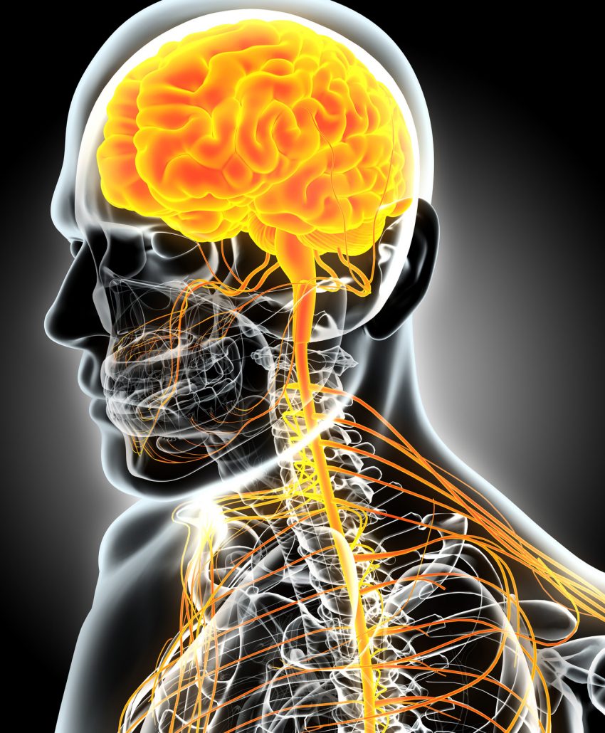
Proprioceptive- Deep Tendon Reflex (P-DTR) is a product of Orthopedic surgeon Dr. Jose Palomar’s original thought and investigations. This work recognizes that sensory receptors (such as those for touch, pressure, hot, cold, pain, etc.) and how the body processes the information from these receptors are paramount in determining neuromuscular responses throughout the entire body.
Motor function is not just determined by the motor system but rather is modified by the inputs from these receptors. These receptors all send information to the brain for processing, and the brain takes this feedback into account when making decisions regarding our movement. If this information is incorrect, as is often the case, the brain is making decisions based on bad information. The result is typically pain and dysfunction. P-DTR uses a comprehensive system of manual muscle testing and neural challenges, involved receptors can be located, and normal, pain-free function can be quickly restored.
Most forms of therapy rely exclusively on addressing the “hardware” of the body, neglecting the fact that much of the pain and dysfunction we experience is often actually a problem with our “software. An example of this would be if your computer has a software bug, changing the monitor or keyboard will not solve it. P-DTR works by effectively identifying, isolating, and eliminating those bugs. The work is rapid and tremendously effective, and unlike anything you’ve experienced before.
Some of the benefits of P-DTR include:
- Maximizes balance and stability throughout the body
- Reduces or ends acute pain in as little as a single session
- Achieves quick, long-lasting results
- Treats the problem rather than the symptoms
- Optimizes overall performance
- Resolves problems you thought you would have to live with
- Accelerates recovery from acute injury
- Increases range of motion, strength, and stamina
- Eliminates lingering dysfunctions and pain from chronic injuries
- Improves muscle function and coordination in a short amount of time
- Does away with the weakened effects of repetitive stress
NeuroKinetic Therapy (NKT)® is well on its way to putting a neurological approach toward healing on the map. David Weinstock’s innovation, NKT®, is a corrective movement system that utilizes specific muscle testing to manipulate the output of information from the Motor Control Center (MCC) in the cerebellum. This part of the brain directs all movement within the body. Like with all other things, the MCC learns through failure. Following some trauma, the MCC adapts to compensatory patterns developed due to injury and pain. Information about when and how to activate different muscles becomes jumbled. The brain reorganizes to carry out still movements that keep the body functional, albeit not efficiently. The body essentially goes into survival mode and continues to proliferate compensation after compensation, even after the initial trauma is perhaps long resolved. These improper movement patterns continue indefinitely unless the MCC is reprogrammed and taught the proper way to move again. NKT® provides this necessary intervention.
NKT® assesses dysfunctions of the MCC via manual muscle testing and corrects faulty movements right from the source. It provides clarity for seemingly irresolvable and nagging issues by supplying a map for the motor output’s inner workings from the brain. Dysfunctional patterns are corrected, and the client is provided with follow-up homework to continue at-home reinforcement of new patterns initiated during the session.
Some of the benefits of NKT® include:
• Utilizes muscle testing to quickly and accurately locate the source of problems
• Fixes faulty wiring in the brain to help achieve results that endure
• Eliminates long-held compensatory patterns developed as a result of old injuries
• Vastly improves the overall efficiency of the body
• Increases muscle coordination, strength, endurance, and range of motion
• Provides clients with at-home correctives to maximize progress
– What is a neurological exam?
A neurological exam also called a neuro exam, evaluates a person’s nervous system. It may be done with instruments, such as lights and/or reflex hammers. It usually does not cause any pain to the patient.
The nervous system consists of the brain, the spinal cord, and the nerves from these areas. There are many aspects to an exam, including assessing motor and sensory skills, balance and coordination, mental status (the patient’s level of awareness and interaction with the environment), reflexes, and functioning of the nerves.
The extent of the exam depends on many factors, including the initial problem that the patient is experiencing, the age of the patient, and the current condition of the patient.
Why is a neurological exam done?
A complete and thorough evaluation of a person’s nervous system is important if there is any reason to think there may be an underlying problem, Following any trauma or to follow the progression of a disease.
If a person has any of the following complaints:
• Headaches
• Blurry vision
• Change in behavior
• Fatigue
• Change in balance or coordination
• Numbness or tingling in the arms or legs
• Decrease in ROM(range of motion)movement of the arms or legs
• Injury to the head, neck, or back
• Slurred speech
• Weakness
• Tremor
Need to add more here ankle sprains, concussions, etc
During the exam, we will test the functioning of the nervous system.
The nervous system consists of the brain, spinal cord, 12 nerves that come from the brain, and the nerves that come from the spinal cord.
• Mental status. Mental status (the patient’s level of awareness and interaction with the environment) may be assessed by conversing with the patient and establishing their awareness of person, place, and time. The person will also be observed for clear speech and making sense while talking. The patient’s healthcare provider usually does this just by observing the patient during normal interactions.
• Motor function and balance. This may be tested by having the patient push and pull against the healthcare provider’s hands with their arms and legs in different directions and motions. Balance may be checked by assessing how the person stands and walks or having the patient stand with their eyes closed while being gently pushed to one side or the other. (Perturbation testing)
• The patient’s joints may also be checked simply by passive (performed by the healthcare provider) and active (performed by the patient and provider) movement.
• Sensory exam The provider may do a sensory test that checks his or her ability to feel. This may be done using different instruments: dull needles, tuning forks, alcohol swabs, or other objects. The provider may touch the patient’s legs, arms, or other parts of the body and have him or she identify the sensation (for example, hot or cold, sharp or dull).
• When you are born, you come with the Newborn and infant reflexes. Different types of reflexes may be tested. In newborns and infants, these reflexes are called infant reflexes (or primitive reflexes) are evaluated. Each of these reflexes disappears at a certain age as the infant grows.
These reflexes include:
• Blinking. An infant will close his or her eyes in response to bright lights.
• Babinski reflex. As the infant’s foot is stroked, the toes will extend upward.
• Crawling. If the infant is placed on his or her stomach, he or she will make crawling motions.
• Moro’s reflex (or startle reflex). A quick change in the infant’s position will cause the infant to throw the arms outward, open the hands, and throw back the head.
• Palmar and plantar grasp. The infant’s fingers or toes will curl around a finger placed in the area.
• Reflexes in the older child and adult. These are usually examined with the use of a reflex hammer. The reflex hammer is used at different points on the body to test numerous reflexes noted by the movement that occurs from the reflex point.
• Evaluation of the nerves of the brain. There are 12 main nerves of the brain, called the cranial nerves. During a complete neurological exam, most of these nerves are evaluated to help determine the functioning of the brain:
• Cranial nerve I (olfactory nerve). This is the nerve of smell. The patient may be asked to identify different smells with his or her eyes closed.
• Cranial nerve II (optic nerve). This nerve carries vision to the brain. A visual test may be given, and the patient’s eye may be examined with a special light.
• Cranial nerve III (oculomotor). This nerve is responsible for pupil size and certain movements of the eye. The patient’s healthcare provider may examine the pupil (the black part of the eye) with light and follow the light in various directions.
• Cranial nerve IV (trochlear nerve). This nerve also helps with the movement of the eyes.
• Cranial nerve V (trigeminal nerve). This nerve allows for many functions, including the ability to feel the face inside the mouth and move the muscles involved with chewing. The patient’s healthcare provider may touch the face at different areas and watch the patient as they bite down.
• Cranial nerve VI (abducens nerve). This nerve helps with the movement of the eyes. The patient may be asked to follow a light or finger to move the eyes.
• Cranial nerve VII (facial nerve). This nerve is responsible for various functions, including the movement of the face muscle and taste. The patient may be asked to identify different tastes (sweet, sour, bitter), asked to smile, move the cheeks, or show the teeth.
• Cranial nerve VIII (acoustic nerve). This nerve is the nerve of hearing. A hearing test may be performed on the patient.
• Cranial nerve IX (glossopharyngeal nerve). This nerve is involved with taste and swallowing. Once again, the patient may be asked to identify different tastes on the back of the tongue. The gag reflex may be tested.
• Cranial nerve X (vagus nerve). This nerve is mainly responsible for swallowing, the gag reflex, some taste, and part of speech. The patient may be asked to swallow.
• Cranial nerve XI (accessory nerve). This nerve is involved in the movement of the shoulders and neck. The patient may be asked to turn his or her head from side to side against mild resistance or shrug the shoulders.
• Cranial nerve XII (hypoglossal nerve). The final cranial nerve is mainly responsible for the movement of the tongue. The patient may be instructed to stick out his or her tongue and speak.
Coordination exam:
• The patient may be asked to walk normally or on a line on the floor.
• The patient may be instructed to do multitasking and tap his or her fingers or foot quickly or touch something, such as his or her nose with eyes closed.

